


Short Answer: Yes.
But unfortunately the process by which teeth heal or “remineralize” is much more complex. And it does not happen as easily or quickly as a blog post says it does. Teeth are made up of hydroxyapatite, a complex crystal structure that contains a large amount of calcium. As bacteria consumes the particles of food and sugar left in your mouth, they release acids. These acids eat away Calcium and other minerals weakening the tooth. This is what is known as caries (cavities). If caught very early on, and minerals are available, from diets high in Calcium, Phosphate, Vitamin D or Fluoride, the crystalline structure can rebuild, reduce sensitivity and sometimes even make the tooth stronger. (See “Is fluoride really toxic?”) These small cavities that are caught early on are called incipient caries. You may have seen some the Blogs and facebook posts on How you can heal your teeth, and that your dentist will never tell you that they can. They often use this quote from a well known dental text.
“It has been shown experimentally and clinically that incipient caries [small cavities] of enamel can remineralize.” – Sturdevant’s Art & Science of Operative Dentistry 4th Edition, 2002.
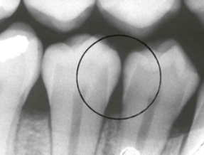
The problem with this is that in the article they avoid explaining what “incipient caries” are. Incipient means “In an initial stage just beginning to happen or develop.” Image A shows incipient or R1 (Grade 1) decay on the premolars. R1 decay or incipient decay is where the decay is only within the outer surface and the halfway point of the full thickness of the enamel. This decay is often missed, but is the decay that can be repaired if the above steps are taken. Because this decay can range from white to dark black in colour. When healed or Calcified the resultant “arrested caries” (Decay that has stopped progressing), can be a variety of colours and if in an aesthetic area the patient may desire a filling anyways.
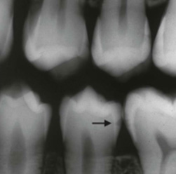
It is important to remember that X-rays do not show all of the extent of the decay and decay can be up to 50% LARGER than it actually appears on the x-ray, So when your dentist sees something like image B, where the decay is about at the 100% through the enamel mark. One can be fairly confident that the decays is already through the enamel. Once that enamel barrier is broken. There is no way for you to properly clean inside the tooth and the decay has entered the softer layers of tissue and will progress much more rapidly. A dentist will normally compare your x-rays with the previous ones so that he can see not only where the decay is at, but also how fast it is progressing. If it is progressing slowly, not at all, or is in the incipient stage he may recommend that you alter your oral hygiene habits in order to halt the growth of the decay. However if the decay is progressing over time, and oral hygiene has not helped, or if the decay is large (see Right) the dentist will recommend some form of repair.
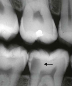
The Decay in image C is R4 (The decay is beyond halfway between the inner wall of the enamel and the pulp chamber where the blood and nerve supply of the teeth reside. If not dealt with soon this could rapidly progress to very painful decay in the pulp of the tooth or an abscess beneath the root, both of which can only be reversed by a Root Canal Treatment or pulling the tooth.